
TumorImager 2TM小动物肿瘤激光扫描测量仪
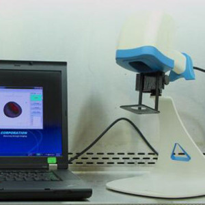
TumorImager 2TM是第二代小动物皮下肿瘤测量仪。TumorImager 2TM用激光扫描技术,对活体小动物皮下肿瘤照射扫描,通过Tumor Manager软件,生成3D图像和数据,并对图像进行综合数据分析。
应用: 小动物皮下肿瘤组织的激光扫描、测量、分析,特别是用在抗肿瘤药物临床前研究,肿瘤进行疾病发生机制和药物筛选的研究。
客户: 肿瘤学及抗肿瘤研究的科研院所、医院、药企,以及为这些单位提供技术服务的企业和模式动物中心等。尤其是开展抗肿瘤药物临床前研究的单位。
TumorImager 2TM与传统卡尺方法对比: 皮下肿瘤移植后,随着它的生长和给药研究,需要多次跟踪测量肿瘤生长状况。传统的小动物皮下肿瘤测量,大都是通过卡尺人工测量、估算的,耗时长,且并极易于产生偏差和错误。Tumor Imager 2采用的激光扫描测量技术迅速、简便,重复性好、误差小,且数据经软件自动存储和分析,不但节约了大量人力,更重要的是提供了一种准确、可信的测量报告, 实时计算体积、TDT、Log Cell Kill, 实时显示图表、曲线; 根据体积、重量等, 提供了30多种方案、数据的报告形式。
产品特点:
* 快速精确测量小动物皮下肿瘤体积,可选择自动和手动肿瘤分割模式
* 自动保存肿瘤图像,进行跟踪和日后分析
* 提高P值准确性和检测的灵敏度
* 激光扫描时间<1秒,实时数据分析时间<2秒
* 几秒内准确测出肿瘤体积
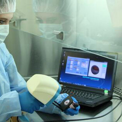
* 小动物不需麻醉,减少劳动成本
* 消除手工测量的主观偏差和记录的错误
* 肿瘤形状独立测量
* 可快速切换手持和支架固定两种模式
* 可准确测量坏死肿瘤
* 真正意义上的高通量测量仪.
技术参数:
* 激光扫描模式:手持和支架
* 肿瘤最大长度:30 mm
* 肿瘤最大高度:20 mm
* 激光扫描时间:<1 sec
* 自动分割时间:~3 sec
* 手动分割模式:触摸屏,鼠标
* 分辨率, Z轴:50μm
* 电脑接口:USB 3.0
* 图像格式:480×640 JPG
* 表面轮廓格式:OBJ,PLY(均为3D模型格式)
* 用户操作方式:触摸屏、鼠标、脚踏板、扫描键
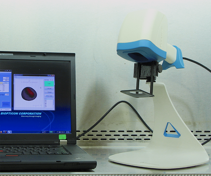
TumorImager 2TM is our next generation tumor scanner for subcutaneous tumor measurements on small lab animals. It uses a unique structured light tumor imaging system to capture both a 3D surface profile and a color image. The result is a one-of-a-kind tumor imaging providing a record of tumor shape and color. The patented algorithms can also isolate a tumor in the recorded image and calculate the volume or area. When interfaced to TumorManager 2TM this leads to a simple but robust, data-verified measurement system that offers speed, accuracy and flexibility.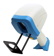
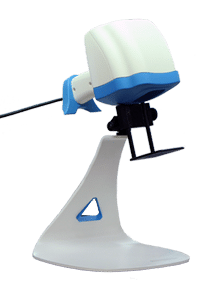
TumorImager 2TM can be quickly converted between a “supermarket-style” hand-held tumor scanner and hands-free scanner using the convenient multi-axis stand. No tools required! The whole unit can be placed in a biosafety hood and easily moved about the lab.
The working principle of tumor imaging
TumorImager 2TM constructs a 3D surface profile by projecting 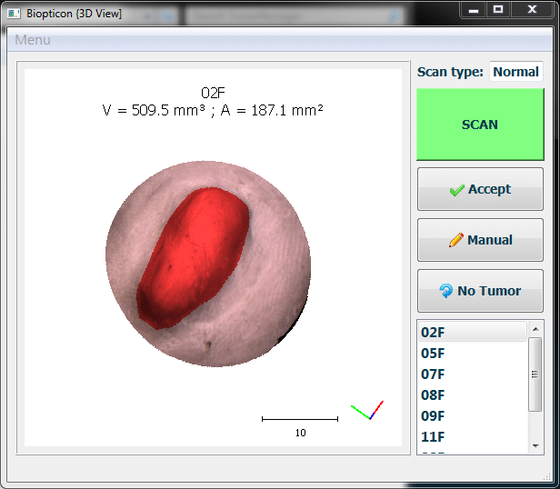 special light patterns on the animal. The relationship between different pattern images allows the creation of an animal surface profile. Like TumorImagerTM, our patented algorithms can automatically isolate a tumor within this profile. Once the tumor is located, volume, area, and other custom statistics are calculated. In addition, a color image and surface profile can be logged for future reference or analysis.
The tumor measurement process
A typical animal scan takes just a few seconds:
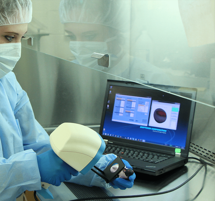
- Position the animal region to be scanned under the black mask plate attached to the scanner. For the hands-free mode, one or two hands can be used to hold the animal.
- Initiate the scan process by pressing the button on the scanner grip, by pressing a foot pedal, or by pressing the scan button on the computer screen.
- After about 1 second, the flashing lights will cease and the animal can be returned to its cage.
- The segmented tumor image together with the tumor volume and area is displayed on the program screen. And the current tumor volume is displayed on the growth curve for this tumor.
- Once the user accepts the scan into the database, the next animal can be scanned.
TumorImager 2TM also easily allows for user-defined regions to be measured and to quickly initiate a rescan. This process generally takes less time than making caliper measurements for advanced tumor imaging.
TumorImager 2TM Specifications
| Scan modes | Hand-held or hands-free stand |
| Maximum tumor size | 30 mm |
| Maximum tumor height | 20 mm |
| Tumor scan time | <1 sec |
| Auto-Segmentation | ~3 sec |
| Manual Segmentation | Touchscreen, mouse |
| Resolution, z axis | 50µm |
| Computer Interface | USB 3.0 |
| Color Imaging format | 480×640 Color JPG |
| Surface profile format | OBJ, PLY |
| User Interfaces | Touch screen, mouse, foot pedal, and scanner button |
System Requirements
- Intel® i5 or better processor
- Microsoft® Windows XP SP3, Vista SP2 or Windows 7, 8, 8.1, 10
- Minimum 4GB of RAM and 750MB of available hard-disk space for program installation
- Minimum 1,280×1,024 monitor resolution video card with OpenGL capability
- Internet or phone connection required for product activation, remote diagnostics, and support
- 1 USB port the TumorImager 2™ and extra USB ports for balance, caliper and RFID readers, etc.
- MS SQL Server 2008 R2, 2012, 2014 or 2016 database